
[ad_1]
A new study published on the preprint server bioRxiv * proposes an integrative computational approach to model the structure of the coronavirus 2 (SARS-CoV-2) envelope of severe acute respiratory syndrome.
Why study the structure of the virus envelope?
One of the different ways that SARS-CoV-2 interacts with the host cell is through its envelope. To get a mechanistic understanding of this interaction, it is important to know details about the structure of the virus envelope. In addition, the surface of the virus envelope offers potential drug targets that can be used to develop future therapeutic strategies.
The challenges of studying the structure of the virus envelope
SARS-CoV-2 belongs to a family of viruses called beta coronaviruses. These are enveloped positive-strand ribonucleic acid (RNA) viruses. Prior to the SARS-CoV-2 pandemic, there were outbreaks of other pathogenic beta coronaviruses, including SARS-CoV and the Middle East Respiratory Syndrome Coronavirus (MERS-CoV).
Therefore, several efforts have been made to elucidate the structural details of various viral proteins and their complexes. Even then, a detailed structure of the entire beta coronavirus particle remains unsolved.
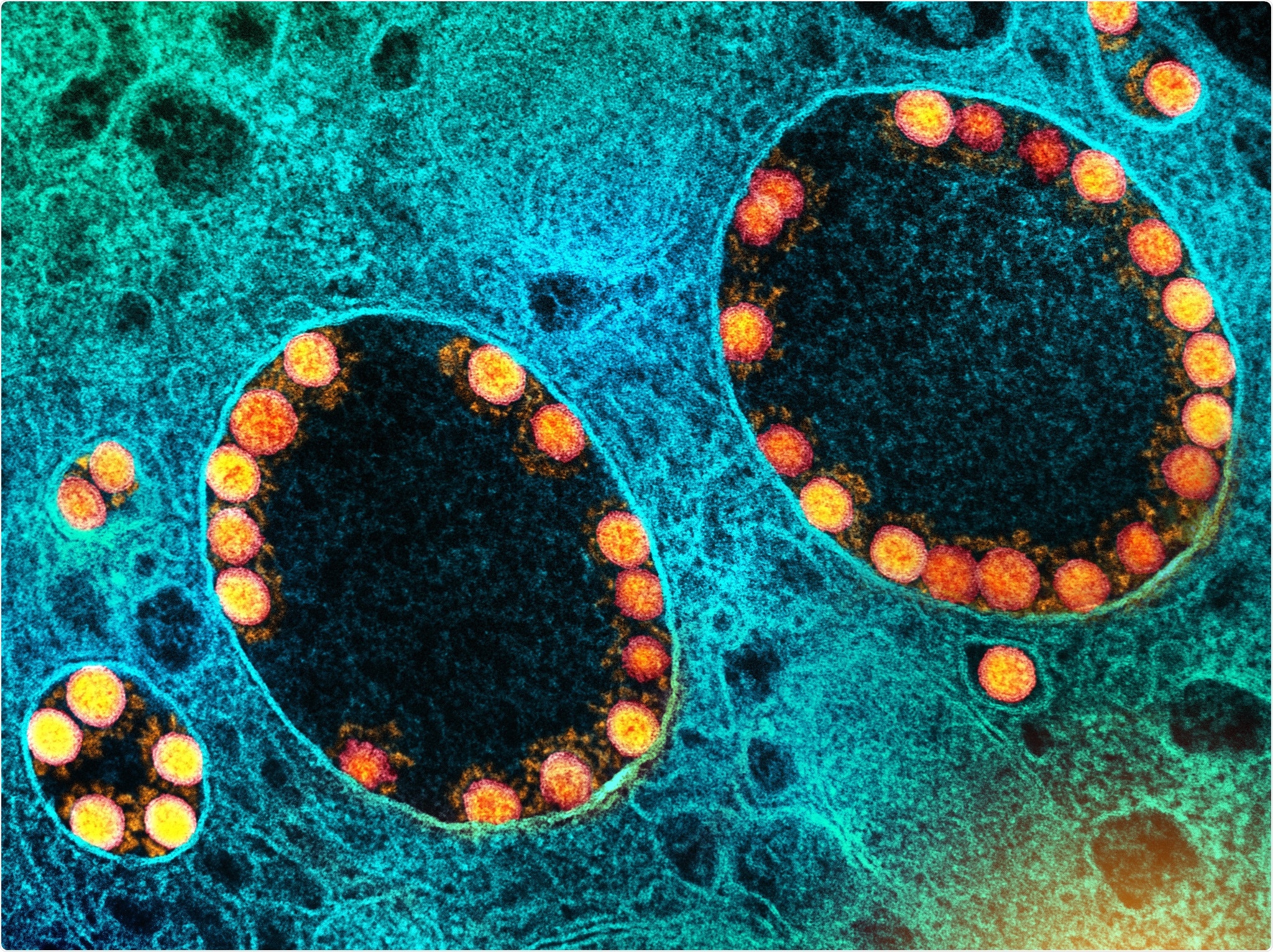
SARS-CoV-2 particles are made up of four structural proteins, including the spike (S) protein, which is required for receptor binding and membrane fusion, the membrane (M) protein, which is essential for membrane construction, the E- Protein, which is present in small amounts, as well as the nucleocapsid (N) protein, which protects the viral RNA. The N protein is responsible for organizing, packaging, and protecting the viral strand of RNA.
In comparison, the S, M, and E proteins come together to form oligomers. The host cell membranes and these oligomers together form the virus envelope.
 Structural characterization of the SARS-CoV-2 virus envelope and its components: A.. An envelope model that was obtained from configuration C1 (2 E-pentamers, 25 S-trimers, 1003 M-dimers) and includes complete structures of S-trimers after 1 μs simulation run. Lipid molecules are shown in sapphire blue, E-pentamers in ruby ​​red, M-dimers in silver, and S-trimers in gold. Major diameters have values ​​of 81.3 nm, 97.8 nm and 103.1 nm. The height of the outer part of the S protein is 25 nm. The surface of the shell shows “filament†patterns, that of transmembrane domains of M dimers are formed while the inner part of the shell shows a close packing of the endodomain arrays of M-dimers; B.. Envelope model from configuration C1 with shortened S-trimer structures at the beginning of the simulations (above) and after 4μs (below); C.. The structural proteins S, E and M, which represent the most important structural components of SARS-CoV-2, wrap themselves in their physiological oligomeric states: S-trimer, E-pentamer and M-dimer. The gray dashed lines correspond to the membrane boundaries. The structures are shown in different scales. D.. Change of the viral shape during the simulation defined by the main gyro radii. The two largest main radii converge towards the value ∼28 nm, while the third converges towards ∼24 nm. The actual diameters of the model after 4μs simulation were 103.1 nm, 97.8 nm and 81.3 nm, respectively.
The virus envelope is flexible and has no symmetry. The structures of the E, M and S proteins have not been established experimentally. There is some structural knowledge of S-trimers and E-pentamers, but no precise model of the M-dimer exists.
In addition, the nature of the interactions between these three proteins is believed to be more complex than originally expected. The complexity and plasticity of the virus make it difficult to study the structure of the virus envelope.
Previous studies
Structural studies of several beta coronaviruses using electron microscopy and tomography have shown that the morphology of the virus envelope is preserved. There is some flexibility in its overall ellipsoidal shape, however.
It is estimated that the SARS-CoV-2 envelope contains 26 ± 15 S-trimers. Although the number of M-dimers in the SARS-CoV-2 envelope is unknown, it is estimated to be around 1,100 based on data from other beta coronaviruses.
Thus, M-dimers are considered to be the most abundant protein in the envelope and their homodimers integrate into the lipid bilayer of the virus. The E protein forms pentamers, which are present in very small numbers on the SARS-CoV-2 envelope.
These studies have provided information about the basic local and global geometric patterns that make up the structural proteins on the shell. However, this information is more general and has a low resolution.
An integrative approach
In the current study, the scientists are pursuing an integrative approach to investigate the virus envelope of SARS-CoV-2. To do this, the researchers characterized S, M and E proteins structurally, both in monomeric and homooligomeric form. In addition, the researchers determined the accumulation of certain lipids due to protein interactions using fingerprinting analysis.
 and structural proteins: E-pentamer (ruby red), M-dimer (silver) and S-trimer (gold). In contrast to the E-pentamer, which remained completely surrounded by the lipid molecules, each S-trimer was supported by at least one M-dimer at the end of the simulation through molecular interaction. Similarly, most M-dimers were found to interact with other M-dimers. B. Two glycosylated S-trimers (dark gray) together with their glycans (orange) after 1 μs simulation run. C. Lipid fingerprinting analysis showed a slight accumulation of negative lipids around M-dimers.")
Protein-lipid interactions in the shell model: A. Local view of the molecular interactions between lipids (sapphire blue) and structural proteins: E-pentamer (ruby red), M-dimer (silver) and S-trimer (gold). In contrast to the E-pentamer, which remained completely surrounded by the lipid molecules, each S-trimer was supported by at least one M-dimer at the end of the simulation through molecular interaction. Similarly, most M-dimers were found to interact with other M-dimers. B. Two glycosylated S-trimers (dark gray) together with their glycans (orange) after 1 μs simulation run. C. Lipid fingerprinting analysis showed a slight accumulation of negative lipids around M-dimers.
In addition, data was collected from multiple sources to compose the initial structural configuration of the shell. The researchers in the current study took data on the refined structures of the three homo-oligomers, their stoichiometry, the composition of the lipid bilayer and the geometry and size of the shell
Molecular dynamics simulations of several configurations were also performed. In addition, a structure and network analysis was carried out in order to reveal characteristic patterns of the structural proteins in the shell arrangement.
Taken together, this approach generated detailed models of the SARS-CoV-2 envelope by building on data from previous studies.
Study results
According to this study, the SARS-CoV-2 M proteins are tightly and stably packed into dimers. The monomers are also complementary in their shape, which enables the natural tiling of several dimers to form “filament†structures. Thus, the M-dimers agglomerate into large, filament-like, macromolecular arrangements that have different molecular patterns.
The results of this computer study are based on previously published experimental data. In addition, the structural and functional insights that this approach provides are outside the scope of data obtained from a single experimental method.
effects
This study provides a basis for expanding our knowledge of the underlying molecular architecture of the entire SARS-CoV-2, the interplay between other protein oligomers, the role of orientation in virus-host interactions, and the molecular mechanisms of virus assembly. In addition, it can help to understand the mechanistic determinants of the stability of the virus particle.
This automated computational protocol can be used to study the envelope structure of other coronaviruses. This structural knowledge can also help in the development of new antiviral compounds as well as virus-like nanoparticles for novel vaccines that can prevent the formation of the virus envelope.
*Important NOTE
bioRxiv publishes preliminary scientific reports that are not peer-reviewed and therefore should not be considered conclusive, that guide clinical practice / health-related behavior or are treated as established information.
[ad_2]